Three-dimensional mapping of intact ovaries reveals the aging dynamics of the ovarian reserve
Abstract
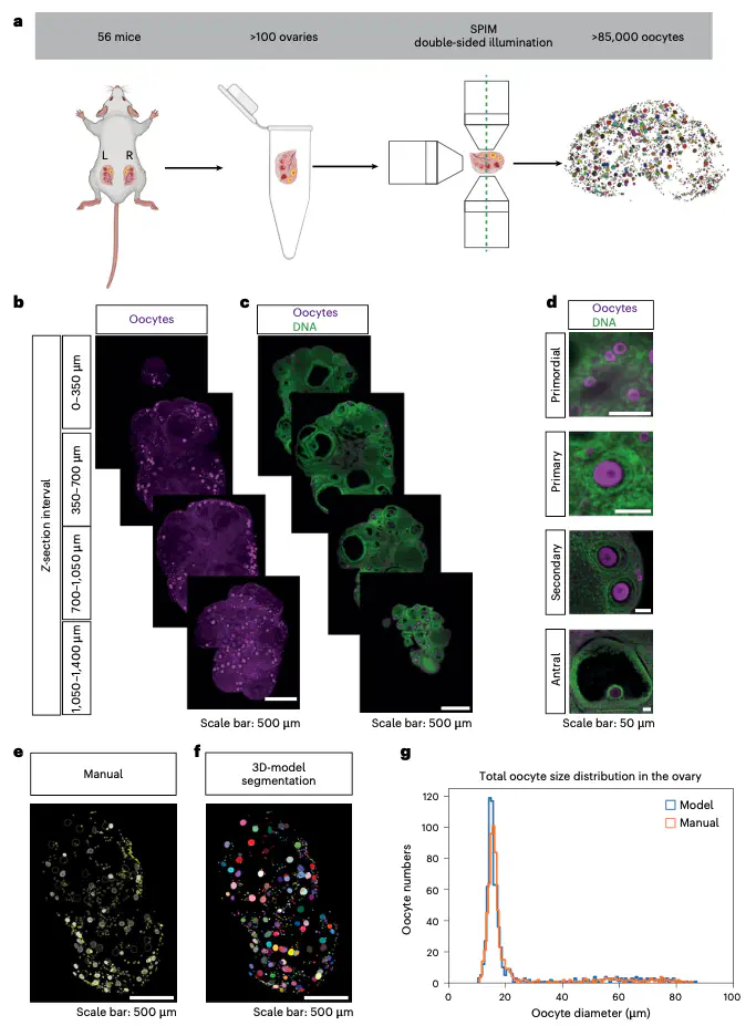
Female fertility depends on a finite pool of oocytes that depletes during aging, yet the spatiotemporal dynamics of this depletion remain poorly understood. Traditional methods obscure the three-dimensional architecture of the ovary, limiting quantitative insights. Here we combine light-sheet microscopy, artificial intelligence-driven segmentation and mathematical modeling to map over 85,000 oocytes in whole ovaries across the reproductive lifespan in mouse. We find that newly activated oocytes represent a fixed fraction of the total oocyte pool despite an age-related decline in oocyte numbers. Spatial analysis revealed that oocytes are enriched along the lateral ovarian axis, and local oocyte density positively correlates with activation. We also uncover a bimodal distribution of oocyte sizes, suggesting a bottleneck during oogenesis. Finally, a differential equation-based model captures the kinetics of oocyte activation and loss. Our findings establish a quantitative framework for understanding ovarian aging and suggest that an organ-scale regulatory mechanism coordinates the age-related decline in oocyte numbers.
Daniel Franco-Barranco
Postdoctoral Researcher
My primary focus is on the development of deep learning solutions for the segmentation of organelles in large-scale and multimodal electron microscopy images.
Ignacio Arganda-Carreras
Ikerbasque Research Associate Professor
My research interests include image processing, computer vision, and deep learning for biomedical applications.